Eye care in the 1800s
It used to be a whole lot worse, as this collection of shocking photos of ophthalmology from the 19th and 20th centuries show.
Published 12 years ago
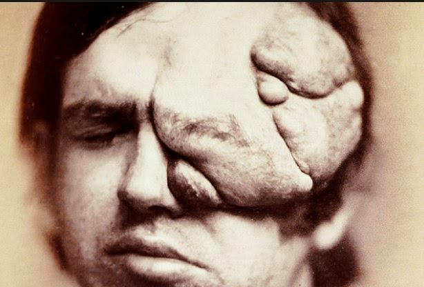
6
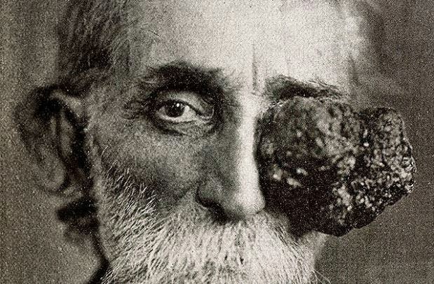
NeurofibromatosisThis photograph first appeared in 1871 in the first medical photographic journal. It shows a patient with von Recklinghausen's disease, a disfiguring hereditary disease now known as neurofibromatosis. There is still no cure for the disease. Patients often have the "fibromas" surgically removed - unless they are too big or too numerous to remove.
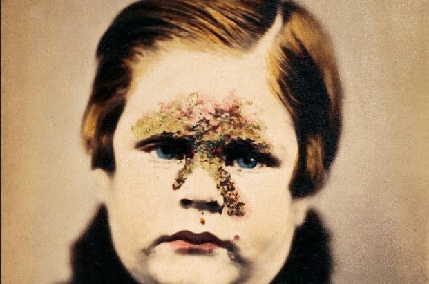
7
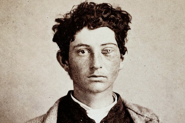
ImpetigoNow rare, the bacterial infection known as impetigo was common in the 19th Century. This photo appeared in 1865 in an English textbook on skin disorders. It shows a child with pustules typical of the disease. Impetigo is typically seen in children, often following a cut, abrasion, or insect bite.
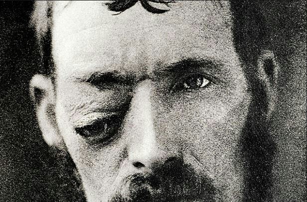
10
Cutting the irisToday's eye surgeons can laser a hole in a patient's iris the colored portion of the eye in a matter of seconds. But in the late 1800s, doctors used a sharp tool to perform "iridectomy," as shown in this 1870 photograph. Iridectomy allowed the creation of an artificial pupil, restoring sight to those blinded by common infectious and inflammatory disorders.
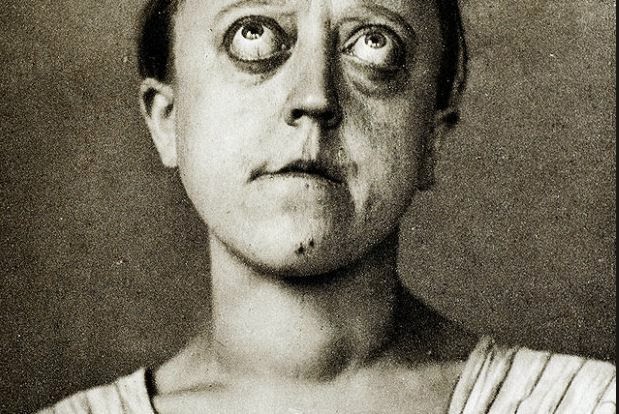
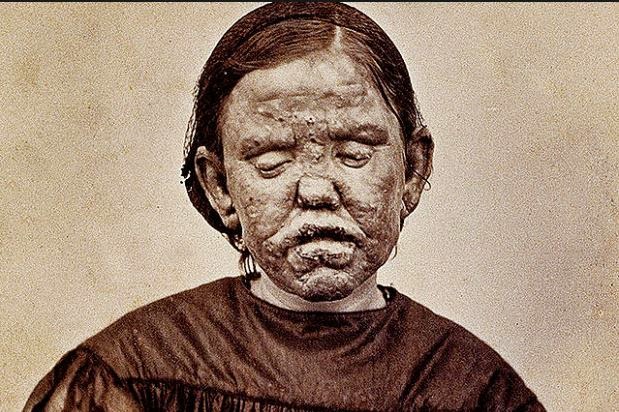
11
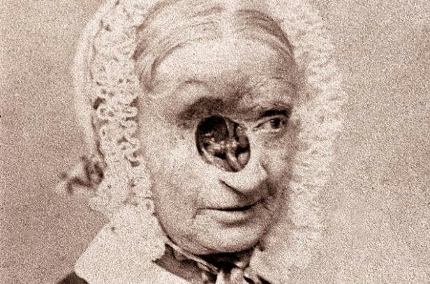
LeprosyThis 1867 photograph shows a woman with a form of leprosy known at the time as "elephantiasis des Grecs." It first appeared in a French medical text published in 1868, "Clinique photographique de l'hospital Saint-Louis." Three years later, in 1871, this dreaded disease - which affects the eyes and causes enlargement of various parts of the body - was found to be caused by a bacterium.
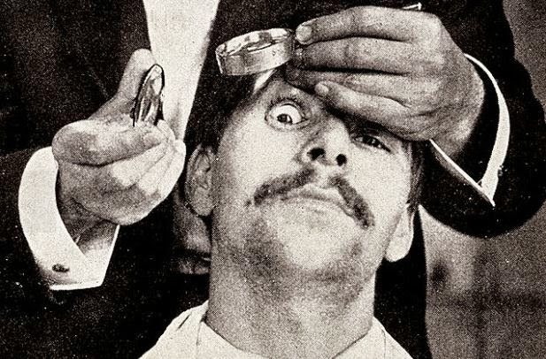
12
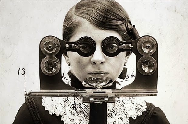
Eye exam by candlelightWhat's the best light source for looking inside the eye? Until the end of the 1920s, when the electric "slit lamp" became widely available, candlelight was the best bet - as shown in this 1910 photograph. Examining the interior of the eye made it possible to conquer many eye diseases.
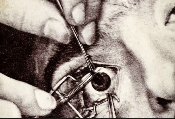
14
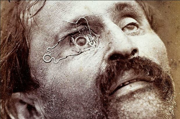
Staged eye surgeryIn 1870, a French ophthalmologist named Edouard Meyer included a series of photos in his textbook on surgery. The film of the era was too "slow" to take photos of actual operations, so he staged photos using cadavers. In this photo, a clamp holds the eye open to show where a scalpel should be positioned to remove a cataract.
Most Popular